 Electrocardiography
Electrocardiography
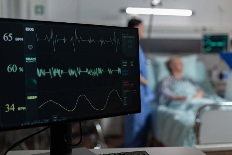
Electrocardiography is a non-invasive medical test used to assess the electrical activity of the heart over time. An electrocardiogram (ECG or EKG) records the heart's rhythm and electrical impulses, providing critical information about heart health. It is one of the most common tests used to diagnose various heart conditions, such as arrhythmias, heart attacks, and other cardiovascular diseases.
Key Components of an ECG:
P Wave: The P wave represents the electrical activity associated with the contraction of the atria (the upper chambers of the heart). It shows how the electrical signal starts in the heart’s sinoatrial (SA) node and spreads across the atria.
QRS Complex: The QRS complex shows the rapid depolarization (electrical activation) of the ventricles, which are the heart’s lower chambers. This part of the ECG corresponds to the contraction of the ventricles that pumps blood throughout the body.
T Wave: The T wave represents the repolarization (recovery) of the ventricles after contraction. It indicates the resetting of the electrical state in the ventricles in preparation for the next heartbeat.
PR Interval: The PR interval measures the time taken for the electrical impulse to travel from the atria to the ventricles. It provides information about the functioning of the heart’s conduction pathways.
ST Segment: The ST segment is the flat line that follows the QRS complex and precedes the T wave. Changes in the ST segment can indicate heart problems like ischemia (restricted blood flow) or a heart attack.
QT Interval: The QT interval measures the time from the start of the QRS complex to the end of the T wave. Prolongation of the QT interval can signal an increased risk of arrhythmias.
Purpose and Clinical Uses of an ECG:
Diagnosing Arrhythmias: An ECG can detect abnormal heart rhythms, such as atrial fibrillation, ventricular tachycardia, or bradycardia. This information helps doctors determine the type and severity of the arrhythmia and plan appropriate treatments.
Detecting Heart Attacks (Myocardial Infarction): An ECG can identify signs of a heart attack by detecting changes in the heart’s electrical activity, particularly in the ST segment and T wave. It can show whether a heart attack is occurring, has occurred recently, or if there are ongoing ischemic issues.
Evaluating Heart Health in Patients with Chest Pain: Patients who experience chest pain often undergo an ECG to assess whether the pain is due to a heart attack or other cardiac conditions. The test can rule out or confirm the presence of heart-related problems.
Monitoring Heart Function in High-Risk Patients: Individuals with conditions such as hypertension, diabetes, or a history of heart disease are often monitored with regular ECGs to check for changes in heart function. This can help prevent complications or worsening heart issues.
Assessing the Effectiveness of Treatments: For patients receiving treatments for heart conditions, such as medications for arrhythmias or pacemaker therapy, an ECG helps evaluate whether the treatment is working effectively.
Identifying Electrolyte Imbalances: An ECG can detect imbalances in electrolytes like potassium and calcium, which are crucial for proper heart function. These imbalances may cause changes in the heart's electrical activity, reflected in the ECG readings.
Pre-surgical and Routine Health Evaluations: ECGs are often part of routine health check-ups, particularly for older adults or those with risk factors for heart disease. The test helps ensure that the heart is functioning properly before surgeries or other medical procedures.
How an ECG is Performed:
Electrode Placement: Small electrodes are placed on the patient’s chest, arms, and legs. These electrodes detect the electrical impulses generated by the heart during its contraction and relaxation phases.
Recording the Heart’s Electrical Activity: The electrodes transmit the heart's electrical signals to the ECG machine, which records them on paper or a digital screen as a series of waves and spikes.
Duration: The test typically lasts only a few minutes and is painless. The patient is usually asked to lie still and breathe normally during the recording to ensure accurate results.
Interpretation: A healthcare professional, typically a cardiologist, interprets the ECG by analyzing the pattern, rhythm, and intervals of the heart's electrical activity to diagnose potential issues.
Types of ECG:
Resting ECG: This is the most common form of ECG, performed while the patient is at rest. It provides a snapshot of the heart’s electrical activity at that moment.
Stress ECG (Exercise ECG): Conducted while the patient exercises on a treadmill or stationary bike, this test helps assess how the heart performs under physical stress. It’s often used to diagnose coronary artery disease or to evaluate exercise tolerance.
Holter Monitoring: A Holter monitor is a portable ECG device worn for 24-48 hours to record the heart’s electrical activity continuously. It is used to diagnose intermittent arrhythmias or monitor heart function over an extended period.
Event Monitor: An event monitor is worn by the patient for several weeks or months. It records the heart’s electrical activity when the patient presses a button, usually when they experience symptoms like palpitations, dizziness, or fainting.
Importance of ECG in Healthcare:
Non-invasive and Quick: ECG is a painless, non-invasive procedure that provides rapid and valuable information about heart health.
Essential for Diagnosing Heart Problems: It is a cornerstone diagnostic tool in cardiology, aiding in the detection of arrhythmias, heart attacks, and other cardiovascular conditions.
Routine Monitoring: ECGs are essential for monitoring patients with known heart disease, those at risk, and those receiving heart-related treatments.
By capturing the heart’s electrical signals, electrocardiography provides critical insights into cardiac health, enabling early detection and intervention for a wide range of heart-related issues.
